Medical/Seismic Imaging Problems Top Research Agenda at IPRPI
October 26, 2004

Figure 1. Arrival time surface (left) and level curves (right), obtained with real laboratory data.
Even today, says Joyce McLaughlin, the Ford Foundation Professor of Mathematical Sciences at Rensselaer Polytechnic Institute, most breast tumors are discovered in an age-old, no-tech way: palpation. "Shouldn't we be able to find an imaging modality that quantifies and extends this exam by detecting abnormal stiffness in tissue?"
McLaughlin, who works in inverse problems, has long followed the literature for signs of progress toward this goal. About three years ago, on reading that Paris experimentalist Mathias Fink had been able to measure the amplitudes of propagating waves, she was impressed enough to seek him out for discussion during a visit with George Papanicolaou at Stanford. After those discussions, her group developed inventive algorithms that worked well on synthetic data. In their current work, they are applying the algorithms to data measured in Fink's lab.
Much of McLaughlin's energy in the last three years has been devoted to this emerging area of inverse problems, called "transient elastography." The goal is to produce diagnostic images of biological tissue that contains abnormal areas of stiffness-mainly, at this point, tumors in the breast or prostate, although she points to other settings, such as tissue damaged by heart attacks, in which the work could apply.
As of this spring, transient elastography is one of several research projects under way at IPRPI-a new center at RPI (with the "IP" standing for Inverse Problems). McLaughlin is IPRPI's director, and, working with RPI mechanical engineer Antoinette Maniatty, she is leading the group in transient elastography; other key members include mathematics postdocs Dan Renzi, Jeong-Rock Yoon, and Jens Klein and grad student Kui Lin, and their counterparts in mechanical engineering, postdoc Larisa Goldmints and grad student Eunyoung Park. This summer, at the SIAM Annual Meeting in Portland, McLaughlin described some of the group's progress in the second AWM-SIAM Sonia Kovalevsky Lecture, "Interior Elastodynamics Inverse Problems: Creating Shear Wave Speed Images of Tissue."
Creative Exploitation of Inventive Experimental Data
For their "innovative hybrid experiments," McLaughlin says, Fink's group in Paris, at the Laboratoire Ondes et Acoustique, Ecole Sup�rieure de Physique et de Chimie Industrielles, created what they call an "ultrafast" ultrasound data-acquisition device. After sending a low-frequency (50-250 Hz) shear wave pulse along a line either on the surface or---"very inventively," McLaughlin says---in the interior of the tissue, the researchers use the device to measure the motion of the slow-moving shear wave that propagates away from this line.
Traditional ultrasound, McLaughlin explains, sends a set of very-high-frequency (4-5 MHz) pulses, recovering the amplitudes of the reflected waves at a rate of about 100 frames/sec. The ultrafast system, by contrast, sends a much larger set of ultrasound pulses, recovering the propagating shear wave amplitudes at about 6000 frames/sec.
Shear waves progagate slowly in tissue because of its high water content, McLaughlin says. At the same time, changes in the elastic properties of the tissue-whether recovered in a palpation exam or in the new hybrid imaging experiments-affect the motion of the shear wave. The high frame rate of the movie of that motion produced by Fink's group allows recovery of the elastic properties. The wave propagates "fast" enough, McLaughlin adds, that only a few milliseconds of data are needed. "This is a real advantage in that the data is not affected by the patient's breathing or other motions."
Useful as this data is, McLaughlin identifies some important features that need to be taken into account. First, the data is noisy: As much as 15% of a measurement can be noise. Second, the data represents only a two-dimensional slice. As the wave propagates, the tissue moves up and down, side to side, and also away from and toward the source, but the experimentalists now measure motion only in one back-and-forth direction.
McLaughlin, Dan Renzi, and Jeong-Rock Yoon have made creative use of this limited data set. Their algorithm exploits the fact that the wave propagates with a front: Ahead of the front, there is no motion; behind the front, the tissue moves back and forth. The front always moves forward in the tissue, and motion in any back-and-forth direction has the same front. This is the basic physical property behind the group's algorithm, which they call the "arrival time algorithm."
The arrival time algorithm recovers changes in shear wave speeds in three steps, beginning with identification of the front locations, or arrival time curves; each curve contains the points at which the front arrives at a fixed time (see Figure 1).
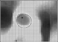
The second step is an initial wave speed reconstruction, based on the changes at successive times of arrival time curves. Simplistically, McLaughlin says, "the speed at any point on a curve is the distance from that point to the next curve, divided by the difference of times associated with each curve. Algorithmically---perhaps surprisingly---it's much faster to take the surface consisting of all the arrival time curves and consider it as the zero level set of a higher-dimensional function." The new function is then extended away from the surface, and the shear wave speed is shown to be a simple first derivative of the new function. "We borrow heavily from the level set community here," McLaughlin says, crediting Dan Renzi "with a major contribution-making the procedure we need for this inverse problem fast." The algorithm concludes with a denoising step. Each of the three steps contributes to the production of images that reveal stiffness changes not seen with ordinary ultrasound (see Figure 2).

Figure 2. With measured lab data, use of the arrival time algorithm reveals stiffness changes not seen with ordinary ultrasound. The circle indicates the approximate location of the inclusion.
"The richness of this problem is becoming more apparent to me all the time," McLaughlin says. The mathematics the group has done to attain these results is new, first published in the journal Inverse Problems in December 2003, and papers now in progress will provide details. As to real-world applications, the group's results to date seem extremely promising. Simultaneously exuberant and cautious, McLaughlin refers to a "testable conjecture" that an extension of their method might be able to distinguish between benign and malignant growths.
New Insight into Active Earthquake Zones
About three years ago, a group of mathematical and geo-scientists and engineers at RPI began a series of joint seminars. The work on transient elastography described in the preceding section had its beginnings in the seminars; a related but significantly different project that also arose, and that also continues within the IPRPI framework, is called "earthquake active-zone fault identification." Leading the work in this area are Steve Roecker, a geo-scientist, mathematical scientists Joyce McLaughlin and Margaret Cheney, and Randolph Franklin, a computer scientist.
Scientists who work on oil deposit location, the researchers explain, start with a man-made surface vibration or explosion and a dense set of seismometers covering the surface in the region of interest; in some cases, they are also lucky enough to have seismometers located along the length of a centrally located borehole. Armed with the data from these sensors, and often using approximate solutions that model only the forward-propagating part of the waves, they draw on a wide collection of algorithms to produce descriptions of the strata of sediments, granites, salt, oil, and ores beneath the earth's surface. Both shear waves and compression waves are recorded at the surface; in this setting, unlike in the human body, the speeds of the two types of waves are reasonably comparable.
Traditionally, earth scientists who study large-scale fault structure in earthquake-prone areas have had much more limited data sets. Because it is so difficult to predict earthquakes, recordings from similarly dense seismic arrays in the vicinity of earthquake events are rare. In the last few years, however, new dense seismic arrays have been positioned near a limited set of very active fault locations. Data from hundreds of earthquakes, many quite small but still providing useful recorded data, have been stored by geo-scientists.
Using only the arrival times of the wave initiated by each earthquake at each seismometer, Roecker has obtained approximate shear and compression wave speed values throughout a large part of an active earthquake region that contains a West Coast fault. This is the start of the new project.
The mathematicians working on the project---McLaughlin, Jeong-Rock Yoon, Alexander Glasman, and Cheney---are now using two separate, but related, ideas to obtain more accurate fault positions. One of these ideas is time reversal-data recorded at the earth's surface is time-reversed, as when a digital tape is reversed. The time-reversed signal then becomes the input to a numerical code that computes the wave as it reverses and propagates back into the earth. At the fault interface, the time-reversed, but now forward-propagating, wave and its reflection either add or cancel. The goal is to use this localized change in the amplitude of the wave to yield a clear delineation of the interface.
The second method utilizes microlocal techniques to approximate the forward-propagating part of the wave, which is strongly influenced by sharp changes in the medium. The goal (one that has already been achieved in the oil-deposit location problem) is to use the changes in wave amplitude to identify interfaces in the medium.
Focal Point for Research in Inverse Problems
Other research activities under way at IPRPI include a broad spectrum of identification problems associated with earthquake dynamics, including bridge embankment integrity and modeling of subduction zones. Center researchers are also investigating radar imaging, along with a variety of additional problems in medical imaging, some based on electrical properties of tissue, and others on x-ray or diffusion tomography.
The center now has nine faculty members, seven postdoctoral fellows, and many graduate and undergraduate students.
An opening conference, held in April 2004, attracted more than a hundred participants. Among the speakers, who addressed a diverse set of topics, were Mathias Fink, Maarten deHoop (Colorado), George Papanicolaou, Erkki Somersalo (Helsinki), Bill Symes (Rice), David Tuch (Massachusetts), and Gunther Uhlmann (Washington). A lively panel discussion, with participants including Bill Rundell, director of the National Science Foundation's Division of Mathematical Sciences, and Art Lerner-Lamm, director of the Lamont Doherty Center for Hazards and Risk Research at Columbia University, discussed maintaining and increasing the scientific pipeline, and future directions and opportunities for IPRPI.

