Next-generation Anatomy Lab
May 3, 2002

Sets of perpendicular planes at each of three points follow Eve's optic nerve up to the "blind spot" at which it touches the retina.
Fred L. Bookstein
"Adam" and "Eve" are pet names for two huge full-color solid images of complete human cadavers. In a technologically strenuous project carried out over the 1990s, one whole solid adult male human and one whole solid adult female human were frozen into blocks of ice and then sliced-every millimeter (Adam) or every third of a millimeter (Eve). Each slice was photographed before it was cut away, and the photographs were scanned at the equivalent of one-third-millimeter object resolution in full color. Adam and Eve are products of the Visible Human Project at the National Library of Medicine.
Images of Adam have been available to the public for some time, and have appeared in books and popular science videos. The images of Eve, all 5700 slices of her, were released more recently.
Our minisymposium, "The Mathematics of Adam and Eve," focused on issues related to the information content of these priceless color images, their effective and speedy transmission from site to site over Internet2, and new ways of viewing the whole solid by exploiting geometric structures more complex than simple slices or shiny surface views.
A high point of the minisymposium was an extended demonstration of Edgewarp, a new program produced by William Green (University of Michigan) and me for the dynamic navigation of continuously tumbling sections of Eve's image. The mathematical model we implemented is the classic "pencil of planes," a continuous one-dimensional family of planes---the set of all the planes normal to a smooth curve, for instance.
The original set of photographs by which Adam and Eve were archived is itself a pencil, although not a very interesting one. Far more intriguing are sets that explicitly follow Eve's curving anatomy. Edgewarp runs on top of a very cleverly packaged version of the full 7-gigabyte volume of Eve generated in a collaboration between the Pittsburgh Supercomputer Center and the University of Michigan.
Dynamic display of sections through this volume are keyed to "filmstrips"---series of separate planes something like preliminary drawings for an animated cartoon. Edgewarp smoothly interpolates a plane-valued function between these slices and then uses each plane in turn to cut a new, artificial section through the actual volume of the full Eve image. On the computer screen or (quite majestically) on the six-foot projection screen, the viewer sees a continuously changing image passing along or across the structure that was used to key the filmstrip, and moving smoothly through the solid Eve at every other point of this section plane.
The displays can be shown three planes at a time---the classic "moving trihedron" of differential geometry---and can be annotated ad lib with the names of organs that are near the planes or that actually cut them. The filmstrips can be run backward or forward at any speed. They can be looked at face---on or tumbled in 3D for a better view, stopped for exploration of particularly interesting regions by hand-control of these pencils, or even "turned sideways," rotated ninety degrees for an alternative movie (e.g., using the osculating plane to a curve instead of the normal).
It is hard to capture these moving images in print, but the three accompanying examples, obtained by coloring in a subset of the keyframes all at the same time in 3D, are an attempt to do so. The first of the three shows a series of planes perpendicular to Eve's corpus callosum (the thick band of neural tissue that connects the two halves of the cortex). The planes that together look like a fanned-out deck of cards go all the way around the curve hinted at in the single plane facing the viewer up the middle. (Visible in the color versions is bright blue material---the latex with which Eve was injected to keep body cavities from falling apart during the sectioning.)
The second example (above) is a series of planes following the optic nerve from about the site of origin of the orbital muscles right up to the "blind spot" at which it touches the retina. This image shows sets of three perpendicular planes at each of three points---notice that the planes are tumbling and rescaling. Finally, the image below is from a site some distance away in Eve's body: the flexor tendon of her right fourth finger, from roughly the mid-forearm through the wrist and out to the corresponding finger. The filmstrip is centered on the narrowest portion of the passage this tendon travels through in the wrist, the exact spot involved in carpal tunnel syndrome.
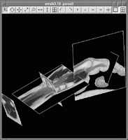
Flexor tendon from Eve's right fourth finger. Centered on the narrowest part of the passage through the wrist, the image vividly illustrates the anatomic changes involved in carpal tunnel syndrome.
The roomful of people who attended this minisymposium were delighted by the navigations of Eve made possible by Edgewarp. We played several of the filmstrips more than once, stopping to show details of anatomical structure or reorienting them as they flew by. For the next SIAM imaging conference, we promised (perhaps rashly) that we would be able to demonstrate filmstrips simultaneously navigating multiple specimens, allowing viewers to appreciate their common structure and their variations at the same time.
The Edgewarp program, a user's manual, and more examples can be found at
ftp://brainmap.med.umich.edu/pub/ewsh3.19.man/.
Fred L. Bookstein is a senior research scientist in the Institute of Gerontology and the Department of Biostatistics at the University of Michigan.
![]()